DISCOGRAM
A discogram is a diagnostic test performed to determine whether a patient's back pain is the result of a spinal-disc abnormality, and, if so, to pinpoint the disc causing the problem. A discogram is performed by injecting a special dye into the patient's spinal disc(s), and using fluoroscopy to view the area in greater detail. The injection creates pressure on the disc and, if the disc is damaged, causes pain.
Due to its invasive nature and the pain it causes, a discogram is used only in instances when a patient has persistent back pain that does not respond to treatment, and to identify damaged discs that need to be removed during spinal-fusion surgery.
The Discogram Procedure
A discogram is usually performed as an outpatient procedure that lasts from about 45 minutes to 2 hours. During a discogram, the patient lies down sideways on a special table. After the patient is properly positioned, the injection site on the back is sterilized. In some cases, an additional anesthetic injection is administered to minimize pain during the procedure.
With the help of fluoroscopy, a needle is then inserted through the skin and into each disc to be examined. After a contrast dye is injected into each disc, the needle is removed. Each disc is then examined with an X-ray or CT scan to see if the dye has traveled; this is diagnostically significant because the contrast dye remains in the center of a healthy disc, but spreads outward in one that is damaged.
Immediately after the discogram, the patient is observed for about an hour. Although it is normal to have some pain at the injection site for a few hours, the physician should be contacted immediately if the pain becomes severe.
A radiologist reviews and interprets the discogram's results, and sends them to the physician. The physician then discusses the results with the patient, and determines an appropriate treatment plan.
Risks of a Discogram
Although a discogram is generally safe, it does carry a risk of complications, which include headache, nausea, allergic reaction to the contrast dye, infection of the area between discs, and injury to blood vessels in and surrounding the spine.
ELECTROMYOGRAM
An electromyogram (EMG) is a diagnostic test that measures the electrical activity of muscles during contraction and at rest. The test is used to determine the cause of muscle weakness, twitches, numbness or paralysis. EMGs help to differentiate symptoms caused by traumatic injury from those caused by neurological disorders.
Reasons for an EMG
When patients are having neuromuscular symptoms, that is symptoms resulting from the connections between nerves and muscles, an EMG is typically administered to diagnose the cause. Diseases and conditions that may be diagnosed through an EMG include:
- Herniated disc
- Amyotrophic lateral sclerosis (ALS)
- Myasthenia gravis (MG)
While an EMG can assist in evaluating problems in the muscles themselves, the underlying causes of such problems may be neurological. The EMG is not able to diagnose diseases of the brain or spinal cord.
The EMG Procedure
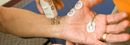
Before the EMG procedure, the areas of skin where needle electrodes are to be placed are well-cleaned. Then very thin needle electrodes are inserted through the patient's skin into the muscle. These electrodes, which detect and transmit electrical signals, are attached by wires to a recording device. After placement of the electrodes, the electrical activity is first recorded with the muscles at rest. Patients are then instructed to contract the affected muscles slowly and steadily and the resulting change in electrical activity is recorded.
The electrodes may be moved to different spots on a muscle, or to different muscles, during the procedure. As the electrodes pick up the electrical activity emitted by the muscles, the information is displayed on a video monitor and recorded. This electrical activity recorded on the monitor provides data about the ability of muscles to respond to nerve stimulation.
Electrical activity in the muscle appears as wavy or spiky lines on the video monitor. There is also an audible representation on the computer speaker of popping sounds when the muscle is contracted. The EMG procedure takes between 30 and 60 minutes. Patients report a sharp pain when the needle electrode is first inserted into the muscle and may experience muscle soreness or tingling for a day or two after the procedure.
While in many cases, an EMG procedure can provide a definitive diagnosis, many patients may have to undergo nerve conduction tests as well.
Risks of an EMG
The EMG procedure is considered safe with very few risks, and complications are rare. There is a small risk of bleeding or nerve injury at the site of needle insertion, though some bruising or swelling is not uncommon. While the needles inserted are sterile, and the chances of infection are minimal, any tenderness, pain, swelling or pus at the site of a needle insertion should be reported to the doctor promptly.
